

 英語
英語 フランス
フランス スペイン
スペイン ロシア
ロシア 韓国
韓国 日本
日本Green Spring Technology's Premium Hyaluronic Acid Enhances Health Product Solutions
ヒアルロン酸 isa natural biopolymer extensively present in numerous tissues throughout the human body。 Since its initial discovery in 1934, hyaluronic acid has become an indispensable, high-value raw material across diverse fields—including biomaterials, premium skincare products, drug delivery systems, and medical care—due to its unique physicochemical properties and exceptional biocompatibility。
Green Spring Technology leverages advanced fermentation processes to provide clients with ヒアルロン酸粉 featuring diverse molecular weight specifications, high purity, and stable performance. We are committed to assisting clients in overcoming core challenges in product development:
・ Exceptional moisturising and rheological properties: Our hyaluronic acid exhibits exceptional hydrophilicity, capable of adsorbing water equivalent to thousands of times its own weight. Its aqueous solution demonstrates outstanding viscoelasticity and strain capacity, empowering clients to develop more comfortable and effective skincare and personal care products.
· Customizable molecular weight selection: Green Spring Technology offers a comprehensive product line ranging from low to high molecular weights, meeting diverse application requirements for material permeability, film-forming properties, and longevity.
・High biocompatibility and safety: Strict quality control ensures strong batch-to-batch consistency, making our products suitable for biomaterials, high-end medical devices, and cosmetic ingredients. This accelerates product launches and enhances market competitiveness.
・Enhanced product innovation: Leveraging its unique non-Newtonian fluid properties and modifiability, Green Spring hyaluronic acid serves as an ideal functional matrix for developing drug delivery systems, post-operative care materials, and premium skincare formulations.
Green Spring Technology empowers clients to achieve product upgrades and technological innovation in the beauty and wellness sector through reliable quality and professional technical support. Should you require hyaluronic acid raw materials, please contact our team to obtain samples and technical documentation.

1. Hyaluronic Acid Raw Materials Drive Innovation in Premium Ophthalmic Health Products
As a natural acidic mucopolysaccharide, hyaluronic acid is gaining prominence as a high-value ingredient in ophthalmic health products due to its exceptional biocompatibility, lubricity, and moisturising properties. Its unique viscoelasticity and pseudoplasticity enable the formation of a gentle protective and lubricating film on surfaces, delivering enhanced comfort to users.
Leveraging its superior physicochemical properties, hyaluronic acid is extensively incorporated into premium eye drops and eye care formulations. It effectively maintains localised moisture levels, enhances product experience, and reinforces surface barrier function. This ingredient provides crucial technical support for the innovative advancement of eye care products.
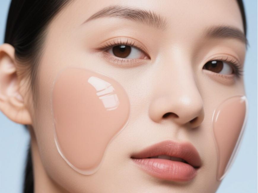
2 . Hyaluronic Acid Ingredients Empowering Innovation in Premium Cosmetic Products
As a key functional ingredient, hyaluronic acid is increasingly utilised in high-end medical aesthetics and skincare products. Its exceptional moisturising properties, outstanding biocompatibility, and controllable rheological characteristics provide crucial technical support for innovation in modern beauty and personal care products.
Naturally present in human skin, this ingredient plays a vital role in maintaining skin structure stability and elasticity. Currently, hyaluronic acid serves as a core component in numerous high-end skincare products—such as moisturisers and hydrating masks—significantly enhancing hydration efficacy and user experience.
Within biomaterials, hyaluronic acid's excellent compatibility and customisable rheological properties make it an ideal choice for developing aesthetically pleasing, stable cosmetic and care products. Simultaneously, this ingredient demonstrates broad application potential in maintaining healthy skin environments and promoting normal metabolism, continuously driving technological innovation and product upgrades within the beauty and personal care industries.
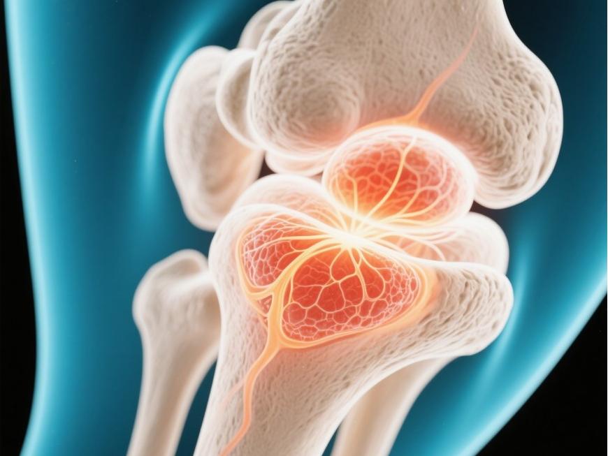
3. Hyaluronic Acid Raw Material Expands Applications in Joint Health Materials
Hyaluronic acid is a key component of synovial fluid, offering excellent lubrication and biomechanical properties that provide effective cushioning and protection for joints. Different molecular weights exhibit distinct rheological behaviours, with high-molecular-weight hyaluronic acid garnering significant attention in joint health products due to its superior viscoelasticity.
Beyond lubrication, hyaluronic acid helps maintain joint internal environment stability and demonstrates excellent biocompatibility in material applications. Hyaluronic acid-based hydrogels modified through cross-linking and other techniques possess superior mouldability and support capabilities. They serve as potential scaffold materials in tissue engineering, providing cells with suitable microenvironments and supporting their growth.
With ongoing advancements in materials technology, innovative applications of hyaluronic acid in joint health and biomaterials continue to expand, offering increased possibilities for product development.
4 . Hyaluronic Acid Demonstrates Broad Potential in Post-Surgical Care Materials
Hyaluronic acid-based biomaterials have garnered significant attention for post-surgical care and tissue isolation applications due to their unique biocompatibility and tunable physical properties. These materials form a gentle barrier layer, providing a supportive environment for wound repair.
Their primary mechanisms of action include: forming a biodegradable bio-protective film on tissue surfaces to provide temporary physical separation; effectively managing exudate during tissue repair to maintain a local moist environment; and supporting normal repair processes by modulating cellular behaviour.
Compared to traditional solid isolation materials, hyaluronic acid-based products offer distinct advantages in application convenience and biocompatibility: Their fluid characteristics enable better adaptation to diverse tissue surfaces, achieving uniform coverage. Furthermore, their in-vivo retention period aligns with tissue repair cycles, eliminating the need for secondary surgical removal.
With ongoing advancements in biomaterial technology, innovative applications of hyaluronic acid in postoperative care are providing novel solutions for product development.
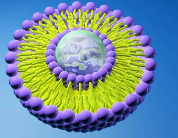
5. The Increasingly Prominent Potential of Hyaluronic Acid in Drug Delivery Systems
As a natural polymeric material, hyaluronic acid demonstrates broad prospects in novel drug delivery system development due to its excellent biocompatibility, modifiability, and biodegradability. Multiple active groups within its molecular structure provide a favourable foundation for drug conjugation and carrier construction.
Through advanced formulation techniques, hyaluronic acid can serve as a functional material for constructing diverse delivery carriers, enabling encapsulation and controlled release of active ingredients. Research indicates that hyaluronic acid-based carrier systems enhance drug encapsulation efficiency, prolong the circulation time of active ingredients within the body, and exhibit certain tissue-selective distribution characteristics.
Furthermore, hyaluronic acid-based carriers can be employed for the delivery of diverse bioactive molecules. By modulating carrier structure and surface properties, effective control over release rates can be achieved. These attributes render it an ideal material choice for developing novel delivery systems, offering fresh avenues for pharmaceutical formulation innovation.
With ongoing advancements in nanotechnology and materials science, hyaluronic acid's applications in drug delivery continue to expand, unlocking greater possibilities for next-generation formulation development.
As a multifunctional biomaterial, hyaluronic acid demonstrates broad application potential across multiple domains. Its unique physicochemical properties and excellent biocompatibility position it as a key ingredient in biomaterials, drug delivery systems, premium skincare products, and post-surgical care formulations.
While expanding its applications, the industry maintains a steadfast focus on material biosafety and application boundaries. Green Spring Technology remains committed to supplying clients with high-quality, highly stable hyaluronic acid raw materials and derivative products. Through rigorous quality control and advanced processes, we ensure product consistency and reliability.
Moving forward, we shall continue collaborating with partners to advance the innovative research, development, and compliant application of hyaluronic acid materials, delivering safer, more efficient raw material solutions across industries.
Green Spring Technology empowers clients to achieve product upgrades and technological innovation within the beauty and wellness sector through reliable quality and professional technical support. Should you require hyaluronic acid raw materials, please contact us at helen@greenspringbio.com or WhatsApp: +86 13649243917 to obtain samples and technical documentation.
参照
[1] meyer k, palmer j w .ガラス質ユーモアの多糖類[j]。1934年(昭和9年)JBiol化学、107(3):629-634。
コーガン[2] G SOLTES L, 厳しいR。 et al. Hyaluronic acid: a natural biopolymer with a broad range of biomedical and industrial applications[J]. Biotechnol Lett,2007,29(1):17-25.
[3]EVANKO S P, TAMMI M I, TAMMI R H, et al. Hyaluronan-dependent pericellular matrix[J]. Adv Drug Deliv Rev,2007, 59(13): 1351-1365。
[4]ローラン T C フレイジャー J R・ Hyaluronan [J]。 FASEB J 1992年には6(7):2397-2404。
【5】黄暁中関国強ヒアルロン酸の生理機能とその応用に関する研究[j]。2015年畜産飼料科学、36(1):25。
[6]タオ国書です。薬理疫学および副作用のモニタリング[j]。chinese journal of geriatrics, 2005(12): 927-928。
[7] girish k s, kemparaju k . the magic glue hyaluronan and its eraser hyaluronidase: a biological overview [j]。^ a b c d e f g h i(2007)、21- 21頁。
[8] chen chao, sun hong, he peihong, et al。フルオロキノロン剤の安全性の違い[j]。^ a b c d e『人事興信録』人事興信録、2004年、6(5)、289-293頁。
-
Prev
Precise Molecular Weight Control: Green Spring Hyaluronic Acid Drives Product Upgrades
-
次
From Skincare to Food: Diverse Applications of Hyaluronic Acid Powder Are Expanding